Cell Structure and Function
What Is the Structure and Function of the Cell Membrane?
The Cell Membrane
All cells are surrounded by a cell membraneglossary term (opens in a new window), which separates the inside of the cell from its surrounding environment. The cell membrane is composed of a double layer of fat molecules called phospholipids. Proteins embedded within this phospholipid bilayer govern which materials move in and out of the cell.
The phospholipids comprising the cell membrane have a distinct molecular structure. Each phospholipid has a head and two tails. The head is hydrophilic, meaning it is attracted to water. The tails are hydrophobic, meaning they are repelled by water. When the phospholipids are exposed to water, the hydrophobic tails of each layer orient toward the interior of the membrane away from the water layer. The hydrophilic heads of each layer orient away from the interior of the membrane, facing either the inside or outside of the cell, toward the surrounding water environment.
Ion Channels and Ion Pumps
Numerous different types of proteins are embedded within the cell membrane. Ion channels are proteins that act like pores in the cell membrane. They control the flow of ions into and out of the cell through passive transport. In passive transport, ions move across the membrane and along their concentration gradient, or from a region of higher concentration to a region of lower concentration, without using chemical energy. In contrast, ion pumps, which are also proteins, transport ions across the cell membrane against their electrochemical gradient or against their natural direction. This process is called active transport, and it requires chemical energy to move the ions.
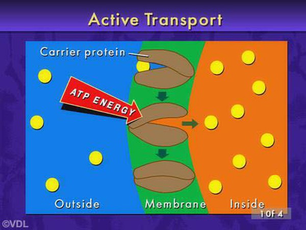
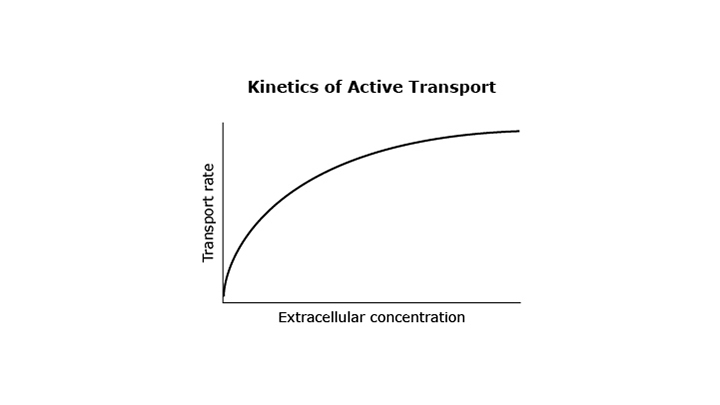
Proteins require energy to move through membranes. What is the difference between passive and active transport?
Teacher Note: Practices
In this item, students explain why a graph of active transport kinetics appear curved. They analyze data using a model in order to make valid and reliable scientific claims. This type of thinking may be challenging to students because it requires them to consider the implications of the active transport model. Support students by discussing the graph’s axis labels and what an increase on each axis means. Extend this item and help students visualize active transport kinetics by building a life-sized model of a cell membrane using large cardboard boxes. Cut three doors to represent three channels in the cell membrane. Time and record data as, one, two, three, and more students enter the cell via the channels. Have students compare the data and evaluate why transport slows down as more and more students try to fit through the three doors. A smaller scale model could use a smaller box with doors sized for smaller objects, such as ping pong balls. The same experiment can be conducted.